More on staining the surface of a dry eye and what it means…What do dry eye tests mean? (Part 4)
Last week I introduced the orange dye called Fluorescein and how it can color the tears in a way that allows dry eye specialists to monitor the “Tear Break Up Time” (TBUT). I mentioned it also stains dry, damaged cells on the surface of the cornea, so a dry eye doctor can assess the damage being done to the ocular surface by this dry and/or dysfunctional tear. This dye is a vital part of most eye exams and the amount, the timing from when applied to when examined and the dilution (and in what product - from anesthetics to salt water) can all have a bearing on how it stains and how it is perceived. There are patterns of uptake that are characteristic of certain eye diseases - so a skilled dry eye doctor has a lot to consider when interpreting how a dye like this reacts with their patient’s eyes. A blue light filter will help the orange dye “fluoresce” where the staining or puddling orange dye will now look bright yellow, to further highlight irregular and damaged areas on the surface of the eye.
Because a cell has to be significantly damaged in order for the dye to be absorbed into the cell (or to stain the footprint of where the cell used to live - but now is gone), the presence of any “staining” can be significant to the exam. Another common stain used to examine a dry eye is Lissamine Green and it is also non-toxic and generally well tolerated by the dry eye patient. It will stain more mildly damaged cells and is taken up by the conjunctiva more than the cornea (where Fluorescein is taken up more by the cornea and less by the conjunctiva), so is often used in conjunction with the orange dye when assessing damage from dry eye disease (which can affect the entire ocular surface). It doesn’t mix as well with anesthetics, but both can be easily diluted with salt water - and can be delivered in an unpreserved cocktail that should not damage any eye - but can give a better overall image of dry eye (amid other causes of) damage.
Where most areas of the body contain discrete focal points of immune fluid drainage and processing centers called lymph nodes, the eye strains and processes immune fluids through the conjunctiva first. Patterns of swelling in that otherwise clear, thin membrane can indicate various likely diseases which are differentiated into two main groups. Papillae are discrete round “bumps” with a small blood vessel at its core and are typically related to allergies and/or bacterial damage where Follicles are discrete round “bumps” of immune cells that push blood vessels away from the center. Fluorescein dye will “pool” around these bumps, highlighting them in a way that makes them more easily identifiable for the doctor to see. The conjunctiva on the underside of the eyelids tends to react more in this way and it is partly for this reason that a dry eye specialist (or any eye doctor) will often “flip over” eyelids (including the larger, upper lids) after applying some dye.
Friction in the area under the upper eyelid margin will cause a “callous-like” reaction (somewhat akin to the guitar-strummer’s thumb strumming the strings). The green dye will help light this up and it is further evidence of a dry eye (and especially of poor oil, since good oil would help lubricate that margin). Since the leading edge of the lid functions like a “windshield wiper” that helps distribute and replenish tears over the eye, the eye doctor term for this callous is “Lid Wiper Epitheliopathy” (or LWE for those in the know - and for more - see below). Friction over the white of the eyeball can lead to “Conjunctivochalasis” (or CCH) and the green dye can also light up zones around the pleats, folds and wrinkles of that now loose membrane while the orange-yellow dye can puddle along these folds (each uniquely highlighting that issue for the doctor to see). Dryness will damage the more exposed membranes and corneal surfaces first, so a pattern of yellow stippled staining over the cornea with the orange/yellow dye and green stippled staining over the conjunctiva will be more prominent in the average dry eye patient’s eyes.
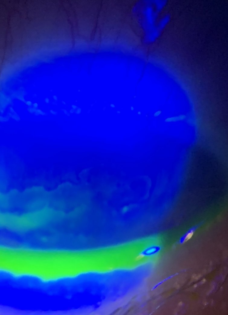
Fluorescein showing “reverse staining” over clear, raised areas of the cornea known as “Map Dot Fingerprint Dystrophy” (lower left side of image where dye pools around and under the irregular patches that don’t stain in this particular patient - see my link on this topic here: https://www.eyethera.com/blog/5b9vbda1r8e8r5y07l9bslgttcl4d8 as well as a few tiny dry spots (small bright yellow dots). The puddle of yellow-green is the tears lying along the lower eyelid margin that shows the typical irregularities or “notching” common to MGD.
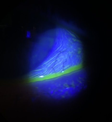
My prior posts on CCH include some of the photos that show how the dye highlights the wrinkles of the otherwise invisible membrane like this one. For a quick link to more on this topic, click here: https://www.eyethera.com/blog/can-conjunctivochalasis-get-better-without-surgery
and here:

This illustration and to learn more at a doctor’s level about lid wiper, see this link: https://www.reviewofoptometry.com/article/lid-wiper-epitheliopathy-what-the-od-needs-to-know
To schedule an appointment with Dr. Jaccoma, call Excellent Vision at either of these two dry eye offices:
(1) 155 Griffin Rd, Portsmouth, NH 03801 (603) 574-2020
(2) 3 Woodland Rd, STE 112 Stoneham, MA 02180 (near Boston) (781) 321-6463