
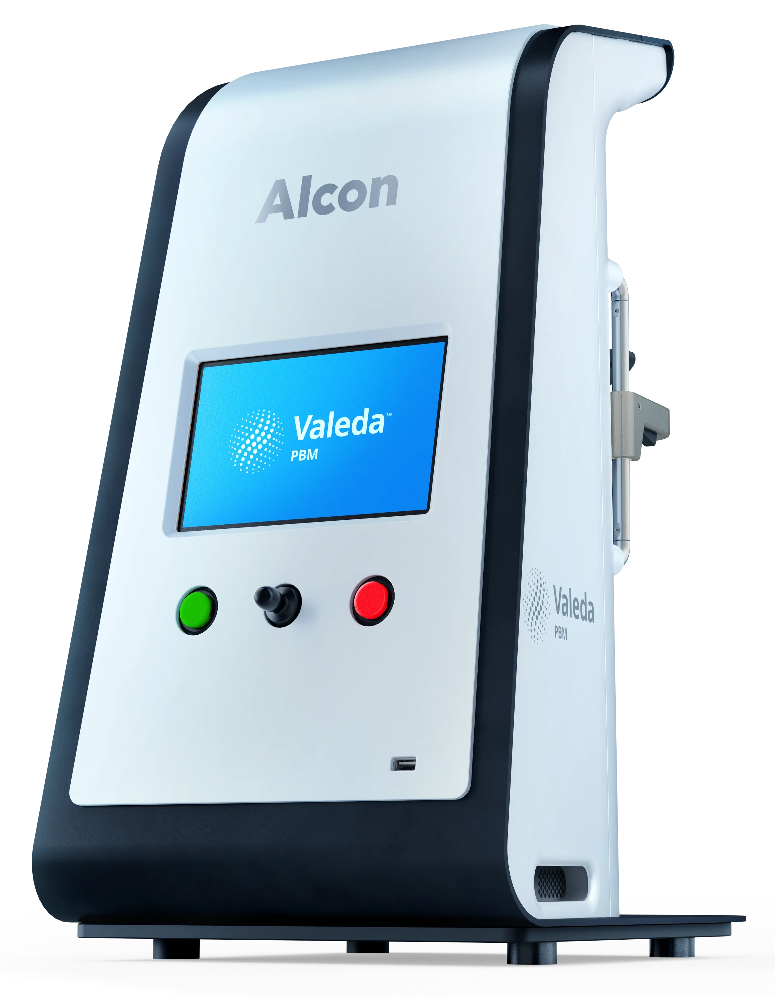
Age tends to lead to a dimming (and sometimes distortion) of vision. Can light therapy “turn back time?” New research suggests this may be true (at least to some extent).
Vision is much more than “seeing.” For many it represents the difference between relying on others for simple tasks vs maintaining their independence. As an eye doctor, I see patients who gradually become restricted from driving, must give up daily tasks such as reading or managing their banking and paying bills and lead to use of magnifiers or techie manipulations of their digital screens to “get by.”
The “Graying of America” is frequently at the heart of this slow, steady, downward spiral of vision and the common issue for seniors is called “Age Related Macular Degeneration,” or ARMD, a term that brings shudders to those who have witnessed this decline of vision in their parents, siblings and/or friends (and can result in the dreaded “shots in the eye” to help maintain that vision).
There are two basic forms of this ARMD. One is called “Dry” and the other is “Wet.” But to understand what is really going on, we need to take a deeper dive.
In the simplest of terms, vision is the result of light entering the eye and striking molecules of light sensitive pigments in the back of the eye. This light sensitive tissue is called the retina and can be divided into a central area (called the macula) and the peripheral area (outside the macula). The central, “macular” area is responsible for the finer vision that can discriminate small letters (measured on eye charts). Once electrons “break off,” they travel through retinal cells to the optic nerve that connects our eyes with our brain and the brain analyses and makes sense of these electrons in the way that generates the “picture” we perceive as sight.
When light strikes, “photochemistry” breaks electrons off of the light sensitive pigments. This leaves a “broken” pigment that must be rejuvenated to remain useful, and this rebuilding requires significant energy and chemistry to occur. Without this rebuilding, the pigments become useless “visual waste” that cannot be effectively removed from the eye. The eye essentially “piles” the debris under the retina, leaving little collections of this waste analogous to the idea that if you could not remove dust from your floor, you might sweep it under a rug. After many years of this janitorial service, the rug would look lumpy and bumpy and as eye doctors, we view these piles as discreet yellowish lumps we call “drusen.”
As drusen accumulate, they raise the retina up like the lumpy rug and when light is cast over these lumps, the straight lines can look crooked or irregular. Typically this occurs slowly, so from one day to the next, most patients don’t routinely perceive it this way, as the brain can adjust the images from each eye to form a more perfect image – but if each eye is carefully judged against the other (commonly with a piece of graph paper with a central dot to hold central focus – called an “Amsler Grid”), then early changes can be more obvious.
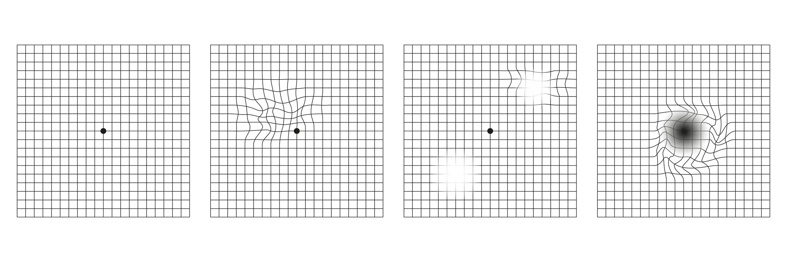
From an AI-generated search, showing a range of common Amsler Grid distortions from ARMD
For more information on the Amsler grid and an excellent educational resource regarding ARMD, see: https://www.macular.org/about-macular-degeneration/what-is-macular-degeneration/monitoring-your-macular-degeneration/amsler-chart
What is also true, is that almost nobody is “perfect” at rebuilding their pigment molecules, so we all tend to create this visual waste as drusen. Some will produce much more than others and certain key factors will contribute to producing more drusen (or in some cases, losing the pigment-bearing cells that aren’t replaced – so called Geographic Atrophy or GA). Some of these risk factors are well delineated in this AI generated list:
Major Non-Modifiable Risk Factors
Age: Risk increases significantly after age 50, with the highest prevalence in adults over 60. [1]
Family History / Genetics: Having a parent or sibling with AMD roughly doubles or triples your personal risk, as inherited gene variations account for a large percentage of cases (such as genes related to the complement system). [1, 2]
Race: Caucasian (white) individuals are diagnosed with AMD at higher rates than other ethnic groups. [1]
Eye Color: People with lighter-colored eyes (blue or green) may face a slightly elevated risk due to lower amounts of protective retinal pigment.
Major Modifiable Risk Factors
Smoking: Cigarette smoking is the strongest preventable risk factor; smokers are up to 3 to 4 times more likely to develop AMD and often experience onset a decade earlier.
High Blood Pressure (Hypertension): Elevated blood pressure places stress on the vascular structures of the eye and increases AMD risk.
Cardiovascular Disease & High Cholesterol: Conditions that affect blood circulation and heart health are closely linked to the development of retinal damage. [1]
Obesity / High BMI: Being overweight or carrying excess abdominal weight raises the likelihood of AMD progression.
Diet: Consistently eating a diet high in saturated fats and low in protective nutrients (such as antioxidants, zinc, and omega-3 fatty acids) contributes to risk. [1, 2] (This is the rationale for the “eye vitamin supplements” such as the scientifically studied and supported Bausch and Lomb “Preservision” formulas II and now III).
Diabetes: Chronically high blood sugar can damage tiny blood vessels in the retina, compounding risk.
Fortunately, the typical drusen we accumulate in our “active” or more independent 60-80 years leaves most of us capable of visually coping. Sadly, there are some who will progress from these little “dry” piles of debris to the more aggressive and more visually threatening form called “Wet ARMD.” Wet ARMD appears to be a reaction of the eye towards dealing with these deposits of visual waste as something to be “healed,” as if it was an injury. This results in sending into the retina “scar tissue” – carried by leaky blood vessels (hence the term “wet”). As the retina absorbs the fluids leaked from these blood vessels, it “swells” and becomes less sensitive – so vision typically becomes distorted (by the irregular surface) and then blurry (as the retina begins to fail from the swelling and from the scar tissue carried into the retina by the blood vessels). Scar tissue is “dumb stuff” that can’t perform the function of the retina. The “shots” commonly administered to slow and stop these leaky blood vessels can stabilize and even reverse early blurring as the blood vessels “regress” and stop leaking. Unfortunately, the drusen that acted as a signal for the blood vessel growth remains and can keep calling them forth – so injections, once started, are frequently required in an ongoing way, to stay the vessels and check the scars.
As an eye doctor helping to counsel my patients who are developing significant piles of drusen and potentially progressing from the dry form to the wet, there has been little to dwell on other than the modifiable risks and monitoring the Amsler grid. Until now. Enter Valeda – a unique form of “Low Level Light Therapy” taking advantage of the “photobiomodulation” I’ve written about as it regards dry eye therapies. More on the specifics of that in my next posting!
To schedule an appointment with Dr. Jaccoma, call Excellent Vision at this dry eye office:
155 Griffin Rd, Portsmouth, NH 03801 (603) 574-2020
PLEASE NOTE – Excellent Vision has CLOSED its office at 3 Woodland Rd, STE 112 Stoneham, MA 02180 (near Boston) (781) 321-6463 and no longer offers any appointments there.